The legalization of cannabis for both medicinal purposes and adult use continues to advance. Consequently, there is a need for simple and reliable analytical methods for the analysis of plant material and cannabis-derived products to support label claims and to ensure quality and safety for consumers.
A robust analytical method has been developed to detect and quantify 18 cannabinoids in cannabis flower, hemp, and edible products using UPLC-PDA and mass detection. Quantitative data were acquired using an ACQUITY PDA Detector combined with the ACQUITY QDa Mass Detector, a single quadrupole mass detector, and controlled by Empower Chromatography Data Software. A Waters ACQUITY UPLC H-Class-PDA-QDa System combined with a CORTECS C18 analytical column enabled the separation of 18 cannabinoids in less than 10 minutes. The lowest level used in the calibration curve was 0.4 mg/mL for cannabinoids detected using QDa mass detection, and the lowest level used in the calibration curve was around 3.125 µg/mL for cannabinoids detected using the PDA. The ACQUITY QDa provided more accurate detection for cannabinoids at low levels compared to the PDA. The results show that this method is suitable for the analysis and accurate detection of cannabinoids across a wide range of matrices including cannabis flower, hemp, concentrates, and edible products.
The legalization of cannabis for both medicinal purposes and adult use continues to advance. As more products are developed and enter the market, the need for simple, reliable, and fast analytical methods to support label claims and ensure quality and safety for consumers grows in importance. The cannabis plant (Cannabis sativa) is a complex natural product that produces hundreds of different cannabinoids, but most laboratories focus their analyses on just five major compounds: Δ9-tetrahydrocannabinol (Δ9-THC), Δ9-tetrahydrocannabinolic acid (THCA), cannabidiolic acid (CBDA), cannabidiol (CBD), and cannabinol (CBN). Recently, other minor cannabinoids have shown beneficial medicinal effects, therefore increasing the importance of separating and identifying them alongside the major compounds. Minimizing peak co-elutions of minor and major cannabinoids in a reasonable timeframe, whilst providing accurate quantitative results, is desirable. This can be particularly challenging in edible products, as cannabinoids have been added to a wide range of foods, including those with high sugar and high fat, like chocolate and gummies. Chemically complex samples require effective sample preparation to ensure successful analysis.1 UltraPerformance Liquid Chromatography (UPLC) in combination with UV detection enables potency determination in complex matrices as it can identify and quantify structurally similar major and minor cannabinoids and their different forms allowing the determination of the correct blend of compounds to provide the most efficacious results. The addition of mass detection can provide both increased specificity and sensitivity. In this study, an ACQUITY UPLC H-Class System with both the ACQUITY PDA and the ACQUITY QDa Mass Detector was used for the analysis of 18 cannabinoids in cannabis flower, hemp, and edible products, including gummies, chocolates, and beverages.
Standard Compounds
Cannabinoid standards were obtained from Cayman Chemical (Ann Arbor, Michigan) and Cerilliant/Sigma-Aldrich (St. Louis, MO).
Reagents
LC-MS grade solvents for sample extractions and LC mobile phases were obtained from Honeywell-Burdick and Jackson (Muskegon, MI). The formic acid was obtained from Sigma-Aldrich (St. Louis, MO).
Cannabis and derived samples were obtained from local sources (Massachusetts). Sample preparation varied depending on matrix.
Cannabis plant material (0.5 g) was weighed into 50 mL centrifuge tubes, and an aliquot of acetonitrile (20 mL) was added along with two stainless steel balls. The samples were processed with a Geno/Grinder® (SPEX, Metuchen, NJ) for 3 minutes at 1500 RPM (Figure 1a). The tubes were sonicated for 20 minutes and centrifuged for 5 minutes at 3000 RCF. After sonication, the samples were centrifuged, filtered through a 0.2 µm PTFE filter. Samples were analyzed in three replicates at two concentration levels at 80x and 2000x dilutions. Some of the main cannabinoids such as THCA and CBDA in the samples are significantly higher and require dilution to bring them into the calibration range.
An aliquot (1 g) of freeze-ground or homogenized samples was weighed out and added to 10 mL water into a 50 mL centrifuge tube. The tube was vortexed for 1 min and sonicated for 20 min. Acetonitrile (10 mL) was added and the tube was vortexed for 1 min. The CEN QuEChERS salts (p/n 186006813) were added to the tube and shaken for 1 min followed by centrifugation for 5 minutes at 3000 RCF. The top acetonitrile layer can be injected directly or diluted with acetonitrile depending on the concentration of the samples.
The Freezer/Mill® was used for grinding gummies and sticky materials; however, embrittled gummy material will become sticky if left at room temperature for extended periods of time (Figure 1b).
An aliquot of the infused beverage (10 mL) was added to 10 mL of acetonitrile. After the sample was vortexed for 1 minute, CEN QuEChERS salts were added, and the sample was shaken for 1 minute and centrifuged for five minutes at 3000 RCF. The top acetonitrile layer can be injected directly or diluted with acetonitrile depending on the concentration of the samples.
Preparation of Pre-spiked Samples:
An aliquot (1000 µL) of a spiking solution containing 18 authentic cannabinoid standards at a concentration of 50 µg/mL was spiked into 1 g of homogenized gummy#941 (predetermined to contain 0.119 % CBD) for a final added concentration of 0.005% in gummy. Water (10 mL) was added and the sample was vortexed and sonicated for 20 min. Acetonitrile (9 mL) was added, the mixture was vortexed and the CEN QuEChERS salts were added. The tubes were shaken for 1 minute and centrifuged for 5 minutes. In the final step, 100 µL of acetonitrile was added to 1 mL acetonitrile extract from the QuEChERS step to make a final volume of 1.1 mL with posts-spiked samples.
Preparation of Post-spiked Samples:
Homogenized gummy#941 (1 g) (predetermined to contain 0.119 % CBD) was added into 10 mL water. The sample was vortexed and sonicated for 20 minutes, followed by the addition of 10 mL of acetonitrile. Next, CEN QuEChERS salts were added. The sample was then shaken for 1 minute and centrifuged for 5 minutes. The final acetonitrile extract (1 mL) was spiked with a mixture containing 18 cannabinoid standards (100 µL of 50 µg/mL).
The concentration of the samples was calculated against a standard curve prepared in solvent and the % recovery of the cannabinoids was determined.
|
LC system: |
ACQUITY UPLC H-Class |
|
Detection: |
PDA single wavelength @ 228 nm, 253 nm PDA Spectrum 210–400 nm at 4.8 nm resolution |
|
Vials: |
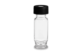
Certified vial (p/n: 186005668CV) |
|
Filter: |
Syringe Filter (p/n: WAT200556) |
|
Column(s): |
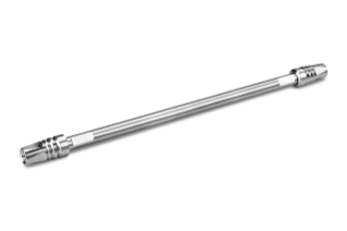
CORTECS C18, 1.6 µm, 2.1 mm x 150 mm (p/n: 186007096) |
|
Column temp.: |
29 °C |
|
Sample temp.: |
5 °C |
|
Injection volume: |
1 μL |
|
Flow rate: |
0.45 mL/min |
|
Mobile phase A: |
20 mM Ammonium formate pH 2.92 |
|
Mobile phase B: |
Acetonitrile |
|
Weak wash: |
90:10, water:methanol |
|
Strong wash: |
5:95, water:acetonitrile |
|
Seal wash: |
90:10, water:methanol |
|
MS system: |
ACQUITY QDa |
|
Ionization mode: |
Positive and negative ion electrospray (ESI+/ESI-) |
|
Acquisition mass range: |
100–600 Da SIR ESI+ and SIR ESI- |
|
Capillary voltage: |
1.5 kV (+), 0.8 kV (-) |
|
Cone voltage: |
10 kV (+), 15 kV (-) |
|
Source temp.: |
150 °C |
|
Probe temp.: |
450 °C |
|
Chromatography software: |
Empower Chromatography Data Software (CDS) |
The analysis of 18 cannabinoids listed in Table 1 was performed using UPLC with PDA and mass detection. The ACQUITY QDa is a robust mass detector designed to be integrated into chromatography workflows for applications that would benefit from mass spectral information. In complex matrices, mass detection can increase confidence in peak identification and can enable lower detection limits.
MS data were collected in full scan ESI positive and negative mode in combination with more specific Selected Ion Recording experiments (SIR). The PDA was set up to monitor individual wavelengths (228 nm and 253 nm) and collect full PDA spectra from 210 to 400 nm. Retention times, PDA data, and mass spectral data together were used to identify detected cannabinoids in the samples (Figure 2). Figure 3 shows the instruments, software, and the analytical workflow that was used to analyze the cannabinoids.
Combining chromatographic, UV and mass data in a single place in the software can ease the burden of data interpretation. The Empower Mass Analysis window (Figure 2) provides a single location to associate chromatographic peaks from all detectors used in the analysis with their corresponding spectra which included the UV chromatogram and spectra are displayed along with the total ion chromatogram (TIC) and mass spectra and extracted ion chromatograms (XIC). Spectra from the detected peaks are time-aligned and displayed in a window above the chromatograms facilitating rapid data review. A chromatogram showing the separation of 18 cannabinoids is displayed in Figure 2, with a total method cycle time of 10 minutes.
Multi-point calibration curves for 18 cannabinoids prepared via serial dilution in acetonitrile were generated and showed good linearity for both PDA and mass detection (R2 > 0.99). The calibration curves ranged from 3.1 to 50 µg/mL for UV data at 228 nm and 0.4 to 50 µg/mL for the ACQUITY QDa. The CBDV calibration curve derived from ACQUITY QDa data had an R2 of 0.9878 due to the saturation of the detector at 50 µg/mL (Figure 4). A calibration range of 0.4–25 µg/mL can be used for some cannabinoids that have a high response with ACQUITY QDa detection and need further dilution to fit the linear curve.
Figure 5 shows a chromatogram of the lowest calibration point used in the study for cannabinoids detected using the PDA at 228 nm and QDa SIR channels. The UV chromatogram shows the detection of the cannabinoids at 3.125 µg/mL with a 1 µL injection. The lowest calibration point for mass detection shown is 0.4 µg/mL.
Using a targeted MS experiment (SIR) allows improved sensitivity and specificity, enabling lower limits of detection in complex matrices. Representative chromatograms from the analysis of low Δ9-THC variety and high Δ9-THC variety cannabis flowers are shown in Figures 6 and 7. In Figure 6, Empower CDS automatically identified and labelled CBD and CBDA in the UV chromatogram based upon retention time. The specificity obtained using targeted analysis is also apparent, increasing the confidence in identified components. Negative ion ESI using SIR targeting m/z 357 was used to detect the acidic cannabinoids, like CBDA. In addition to the target SIR and extracted wavelength chromatograms, full scan MS data and PDA spectra from 210–400 nm were simultaneously recorded allowing the MS and UV spectra to be viewed and thus further increasing confidence in the identified peaks.
Similarly, in Figure 7, the Empower CDS identified and tagged Δ9-THC and THCA in the UV chromatogram based on the retention times. The SIR channels of m/z 315 and m/z 357 show the SIR chromatograms for Δ9-THC and THCA.
An Empower report showing the results from the analysis of a low Δ9-THC variety cannabis sample is shown in Figure 8. The calculated concentrations are displayed in the table in both µg/mL and also weight percent (wt%). Custom calculations can be designed to automatically calculate and report total THC and total CBD, as well as CBD/THC ratio. This reduces the need to perform the calculations separately and keeps the data in a single software environment. The Empower report is completely customizable to report relevant data.
The quantitative results generated from the analysis of 18 cannabinoids in cannabis flower and hemp are shown in Table 2 and were calculated by wt%. Samples were from different manufacturers that contained various types and concentrations of cannabinoids. Samples with high THCA or CBDA were diluted 2000x to bring the samples within the calibration range. Minor cannabinoids at lower concentrations required less dilution (80x dilution). Most of the detected amounts were between 53% to 121% compared to values on the label claim. Some cannabinoids had high %RSD due to the low signal in the detector.
Table 2. Quantitative results from the analysis of cannabis and hemp flower.
%wt1: % weight detected
%RSD: Relative Standard Deviation (n=3)
% Label: % Detected amount compared to amount Label claimed
%wt2: % weight on Label
The quantitative results for the cannabis edibles and cannabinoid infused beverages are shown in tables 3a and 3b respectively. The cannabinoids detected in edibles and infused beverages were calculated by %. Most of the detected amounts of cannabinoids ranged from 60–125% of label claim, except for gummy #868, where the detected value was 36% of the label claim.
Table 3a. Quantitative results from the analysis of edible products.
%wt1: % weight detected
%RSD: Relative Standard Deviation (n=3) *%RSD by QDa
% Label: % Detected amount compared to amount Label claimed
%wt2: % weight on Label
Table 3b. Quantitative results from the analysis of infused drinks. Only CBD was detected in these samples.
%wt1: % weight detected
%RSD: Relative Standard Deviation (n=3)
% Label: % Detected amount compared to amount listed on label
%wt2: % weight on label
Figure 9 shows representative chromatograms from the analysis of cannabinoids in chocolate sample #515. Δ9-THC showed a high UV and QDa response. The signal for Δ8-THC was less for UV compared to QDa, resulting in a %RSD of 28% when analyzed by UV and 0% when analyzed by QDa. MS analysis increased the confidence of identification of Δ9-THC and Δ8-THC in the chocolate sample.
Quantitative results for the % recoveries of spiked cannabinoids in edibles are shown in Figure 10. Recoveries were calculated by comparing the wt% for samples spiked prior to QuEChERS extraction (pre-spiked samples) with the wt% for samples spiked after QuEChERS extraction (post-spiked samples). The concentration of the spiked gummy at the 0.005% level (5 mg/mL in the samples) is close to the detection limit of the calibration curve in the UV analysis (3.15 µg/mL). This could affect recoveries of cannabinoids in some of the matrices at this level due to matrix effects. The recoveries of cannabinoids analyzed with the PDA ranged from 60% to 125%. The recoveries for cannabinoids analyzed with an ACQUITY QDa Mass Detector ranged from 94% to 106%, indicating improved accuracy over UV detection methods.
A comparison of the detector response obtained for the cannabinoids using the PDA at 228 nm and the SIR channels is shown in Figure 11. The detector response for the cannabinoid mixture spiked at a level of 5 μg/mL in the gummy matrix is higher using the MS which can aid in quantifying cannabinoids at lower levels in cannabinoid-infused products.
The Waters ACQUITY UPLC H-CLASS-PDA-QDa System combined with the CORTECS C18 Column enabled effective separation of 18 cannabinoids in under 10 minutes.
The ACQUITY QDa Mass Detector provided orthogonal detection to PDA for the confirmation of peak identities and quantitation of cannabinoids at low levels. The lowest level used in the calibration curve was 0.4 µg/mL for cannabinoids using mass detection, and the lowest level used in the calibration curve was around 3.125 µg/mL for cannabinoids using PDA.
The Empower Mass Analysis window provides a single location to associate the chromatograms and spectra from all detectors used in the analysis. The consolidation of this information in one place makes data review and interpretation easier to manage.
Empower CDS has numerous features which help in analysis of the data including tailored calculations allow relevant information to be derived quickly. Performing the calculations within the software maintains integrity and aids in record keeping.
QuEChERS extraction is effective for extraction of cannabinoids in edibles and drinks with high recoveries.
This analytical workflow is suitable for the analysis of cannabinoids across a wide range of matrices including cannabis flower, hemp, concentrates and edibles, and beverages.


720007199, March 2021